Case 20
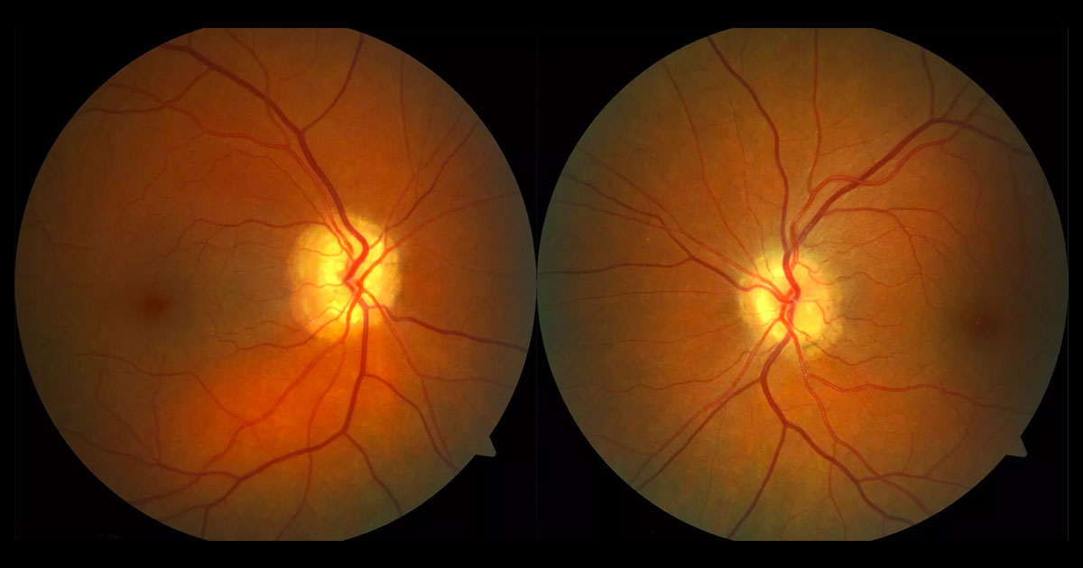
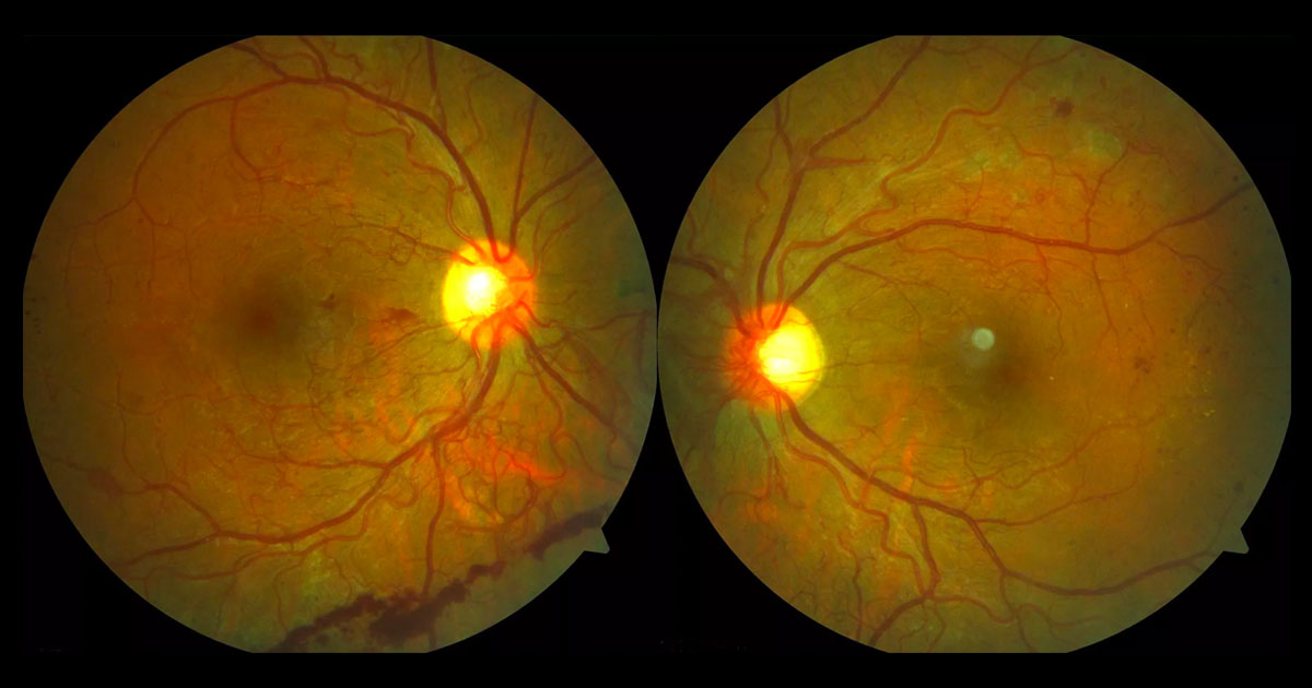
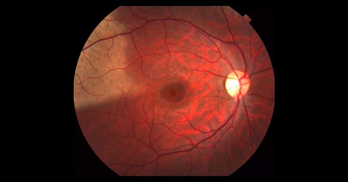
A 46-year-old lady was referred by her optometrist with an abnormal optic disc appearance and flashes in her right eye.
Here you’ll find interesting cases of eye conditions along with news and developments in the ophthalmology world.
Cases are presented as an initial image with history and examination. Health practitioners are encouraged to deduce the condition, before further investigations, diagnosis and management are presented.
We hope you find it as educational, informative and exciting as we do!
Click here to view our newsletter privacy notice.
The information provided during signup is used by Eye Specialists Centre to send newsletters using the cloud-based software, Mailchimp. We do not disclose or share your personal data with other third party without your consent, or unless it is required by law. If you have any concerns about your privacy, please do not hesitate to ask.
A 46-year-old lady was referred by her optometrist with an abnormal optic disc appearance and flashes in her right eye.
Tags: flashes, prominent optic disc, raised intracranial pressure, optic disc drusen
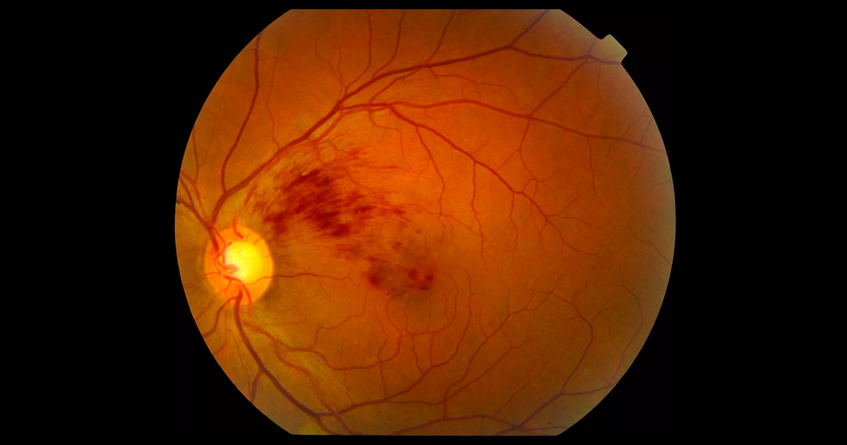
A 22-year-old man presented with a “blob” in the centre of his right vision.
Tags: preretinal haemorrhage, diabetic retinopathy, valsalva retinopathy
A 10-year-old boy was referred with blurred left vision following blunt ocular trauma.
Tags: trauma, vitreomacular traction syndrome, traumatic full-thickness macular hole
A 29-year-old woman was referred with blurred vision and flashes in her left eye.
Tags: blurred vision, flashes, acute multifocal placoid pigment epitheliopathy, multiple evanescent white dot syndrome
A 22-year-old mother was referred after noticing new floaters in her right eye.
Tags: floaters, pregnancy, diabetic retinopathy, neovascularisation
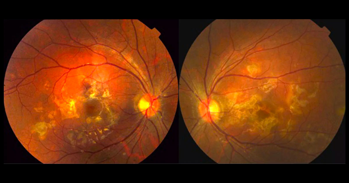
A 54-year-old Caucasian female saw her optometrist complaining of bilateral flashes and floaters.
Tags: floaters, flashes, sarcoidosis, birdshot chorioretinopathy
A 13-year-old girl was referred with a three month history of seeing “black spots” in her left vision.
A 39-year-old man was referred with gradual loss of vision in both eyes noted over the last six months.
Tags: gradual loss of vision, yellow foveal spots, vitreomacular traction syndrome, alkyl nitrite popper retinopathy
A 28-year-old Asian male was referred by his optometrist after being hit in his right eye by a basketball.
Tags: trauma, retinal whitening, infective retinitis, commotio retinae
A 42-year-old Caucasian female was referred by her optometrist with a 1 week history of mildly reduced vision in her left eye.
Tags: reduced vision, branch retinal vein occlusion, macular oedema
A 21-year-old male was referred with acute bilateral loss of central vision.
Tags: bilateral vision loss, photophobia, acute posterior multifocal placoid pigment epitheliopathy, serpiginous choroiditis
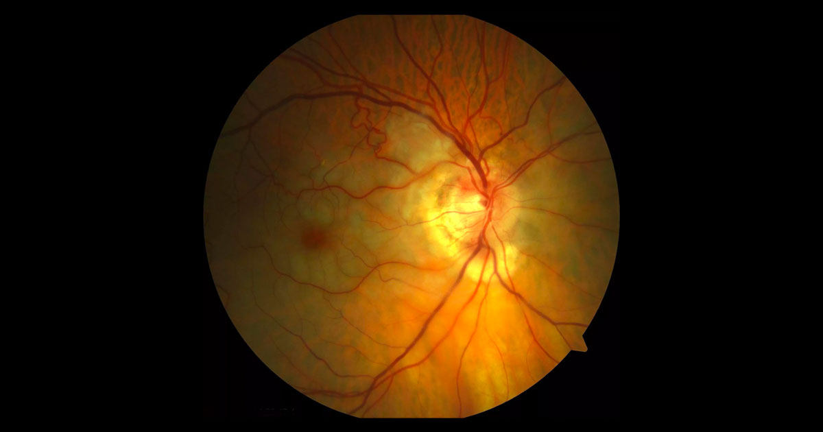
A 76-year-old man was referred with acute painless visual loss in his right eye.
Tags: painless vision loss, central retinal vein occlusion, central retinal artery occlusion, anterior ischaemic optic neuropathy
Have a question? Call one of our clinics today.
© 2019-2024 Eye Specialists Centre | Privacy Policy | Disclaimer | Website design: ![]()