
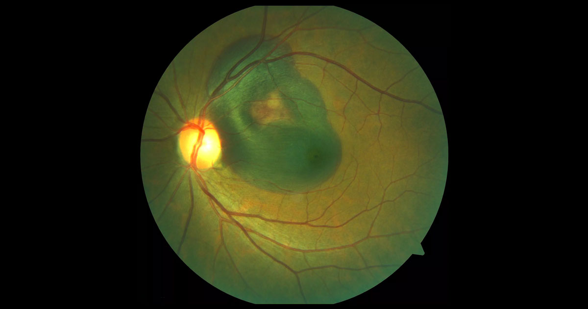
Case 32
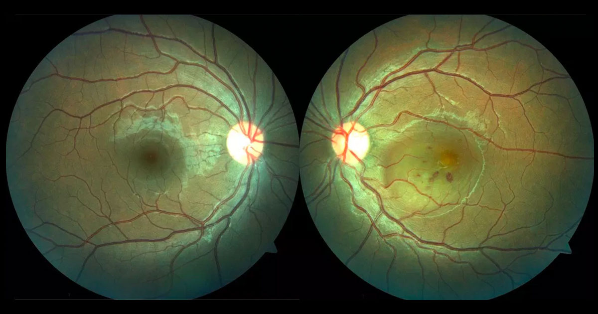
A 43-year-old female was referred with a possible right retinal detachment.
Here you’ll find interesting cases of eye conditions along with news and developments in the ophthalmology world.
Cases are presented as an initial image with history and examination. Health practitioners are encouraged to deduce the condition, before further investigations, diagnosis and management are presented.
We hope you find it as educational, informative and exciting as we do!
Click here to view our newsletter privacy notice.
The information provided during signup is used by Eye Specialists Centre to send newsletters using the cloud-based software, Mailchimp. We do not disclose or share your personal data with other third party without your consent, or unless it is required by law. If you have any concerns about your privacy, please do not hesitate to ask.
A 43-year-old female was referred with a possible right retinal detachment.
Tags: headache, floaters, retinal detachment, degenerative retinoschisis
A 20-year-old man was referred with a central scotoma in his left eye.
Tags: scotoma, retinal detachment, choroidal neovascularisation, acute idiopathyic maculopathy
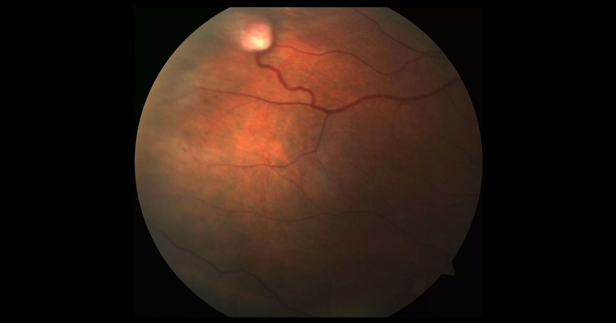
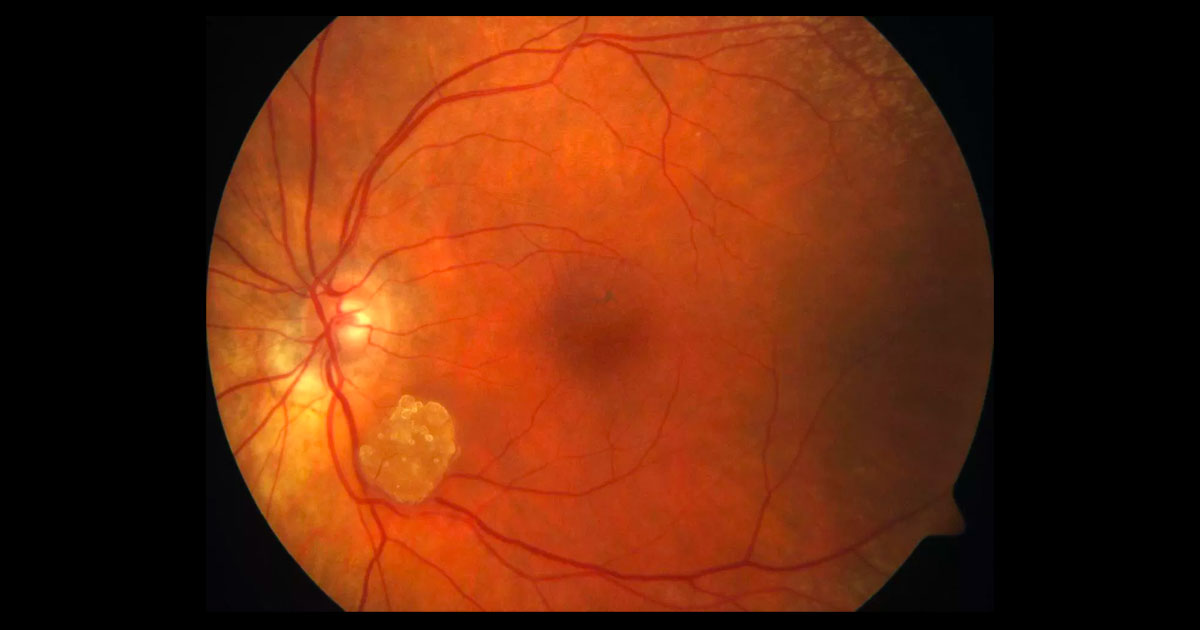
A 33-year-old female was referred with a right supero-temporal retinal lesion.
Tags: retinal lesion, retinal capillary haemangioblastoma, von Hippel-Lindau, vasoproliferative tumour
A 24-year-old female engineer presented with a 1 week history of bilateral blurred vision with paracentral scotomas and occasional flashes.
Tags: blurred vision, scotoma, flashes, acute posterior multiple placoid pigment epitheliopathy
A 60-year-old female was referred with an incidental finding at her left macula.
Tags: vitreous detachment, lamellar macular hole, macular psedohole
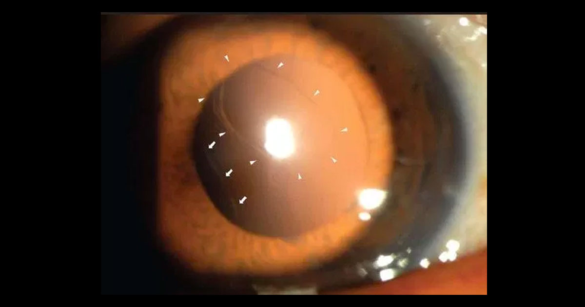
A 72-year-old male was referred with reduced vision in his right eye following cataract surgery.
Tags: reduced vision, cataract surgery, diabetic macular oedema, pseudophakic cystoid macular oedema
A 91-year old lady was referred by another ophthalmologist with a mass in her left eye.
Tags: preretinal mass, granuloma, retinal astrocytic hamartoma, retinal gliosis
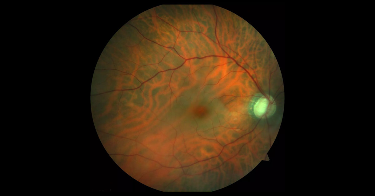
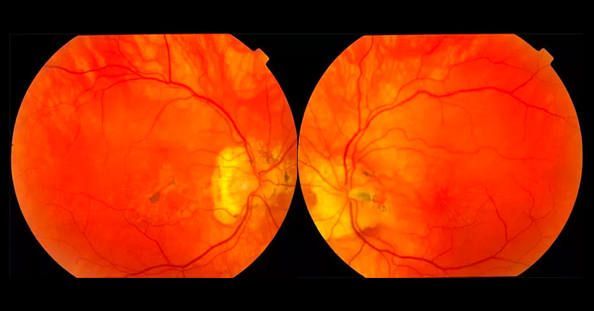
Figure 1. Colour photograph of the right fundus at presentation demonstrates retinal pallor with a cherry red spot at the macula. The cup disc ratio is enlarged.
A 68-year old man was referred with profound vision loss one day following cataract surgery.
Tags: vision loss, cataract surgery, central retinal artery occlusion, retinal toxicity
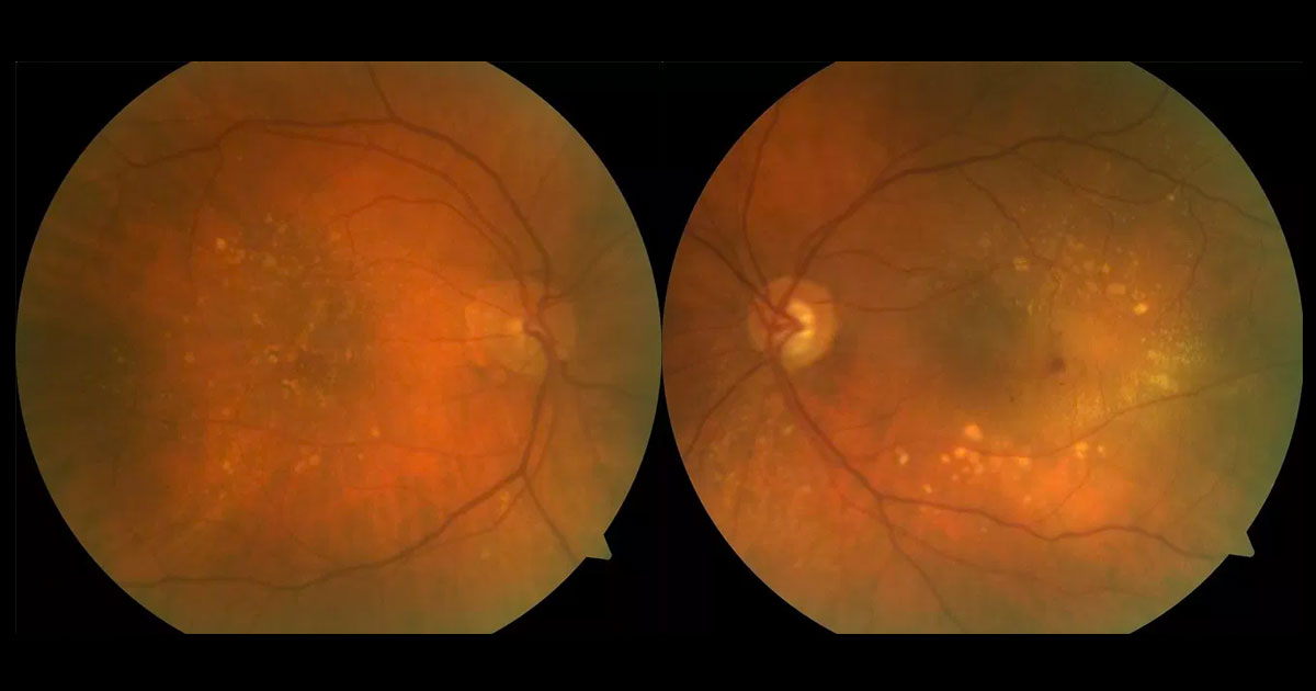
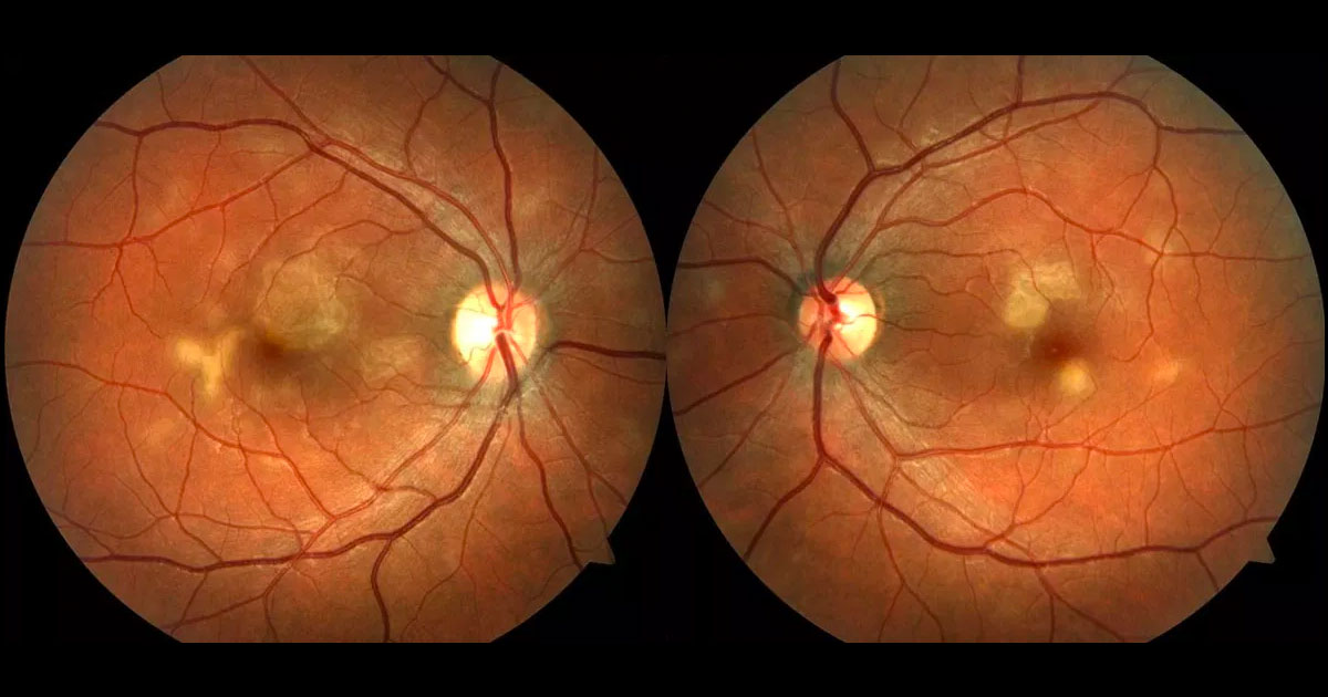
A 66-year old female was referred with blurred vision in her left eye.
Tags: blurred vision, intraretinal haemorrhage, age-related macular degeneration, diabetic retinopathy

A 37-year old lady was referred from an optometrist with a two-week history of mild frontal headache and transient loss of vision.
Tags: swollen optic disc, raised intracranial pressure, space occupying lesion, idiopathic intracranial hypertension, reduced vision
A 59-year-old female was referred with acute painless loss of central vision in her left eye.
Tags: reduced vision, submacular haemorrhage, age-related macular degeneration, choroidal neovascularisation, polypoidal choroidal vasculopathy
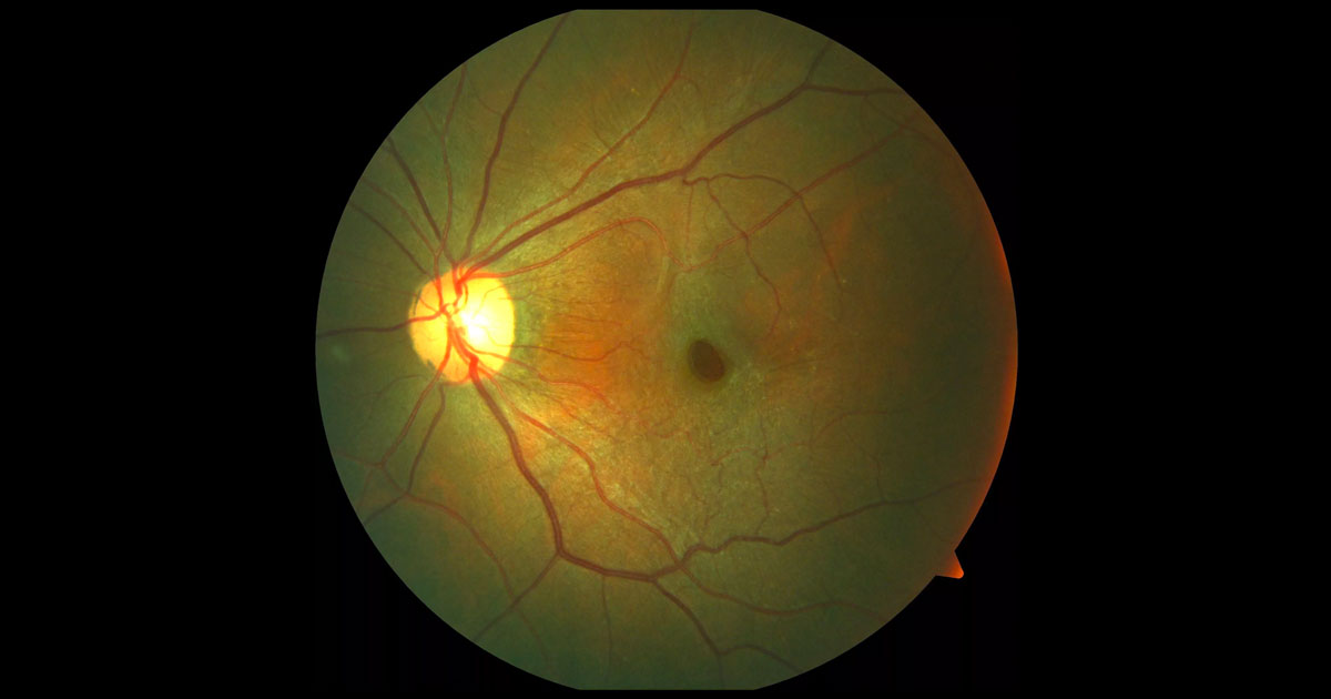
A 41-year-old male was referred with distortion in his right eye.
Tags: drusenoid deposits, age-related macular degeneration, familial dominant drusen
Have a question? Call one of our clinics today.
© 2019-2024 Eye Specialists Centre | Privacy Policy | Disclaimer | Website design: ![]()