Case 44
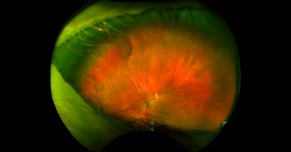
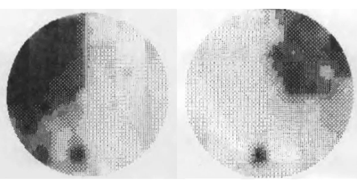
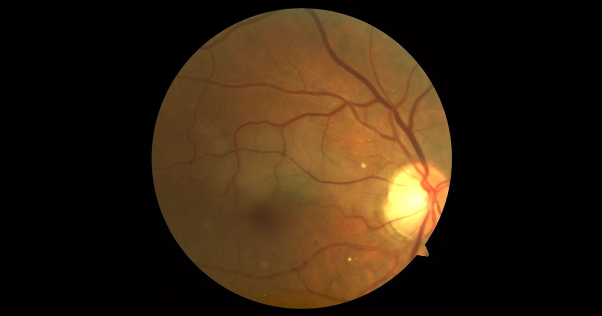
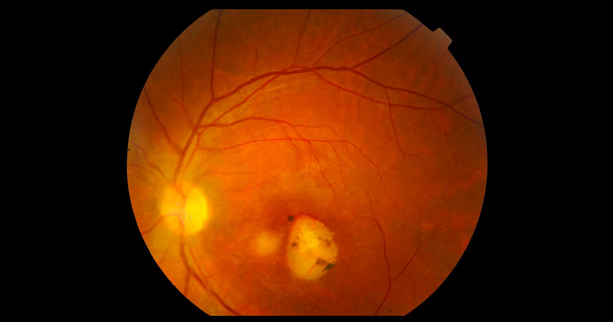
A 79-year-old woman was referred by her general ophthalmologist with left pseudophakic cystoid macular oedema and a raised peripheral sub retinal lesion.
Here you’ll find interesting cases of eye conditions along with news and developments in the ophthalmology world.
Cases are presented as an initial image with history and examination. Health practitioners are encouraged to deduce the condition, before further investigations, diagnosis and management are presented.
We hope you find it as educational, informative and exciting as we do!
Click here to view our newsletter privacy notice.
The information provided during signup is used by Eye Specialists Centre to send newsletters using the cloud-based software, Mailchimp. We do not disclose or share your personal data with other third party without your consent, or unless it is required by law. If you have any concerns about your privacy, please do not hesitate to ask.
A 79-year-old woman was referred by her general ophthalmologist with left pseudophakic cystoid macular oedema and a raised peripheral sub retinal lesion.
Tags: macula oedema, subretinal lesion, choroidal naevus, vortex vein varix

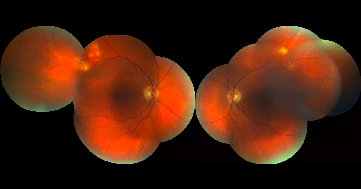
A 34 year male was referred with blurred vision in his left eye.
Tags: blurred vision, unilateral cataract, trauma, fuch's hereochromic iritis, retinal detachment
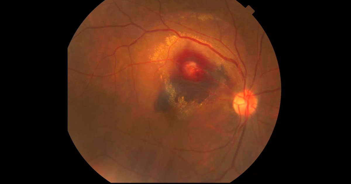
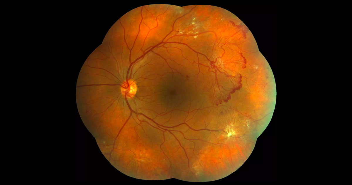
A 72-year-old woman was referred with acute painless vision loss in her right eye.
Tags: painless vision loss, choroidal neovascularisation, ruptured retinal macroaneurysm, Terson's syndrome
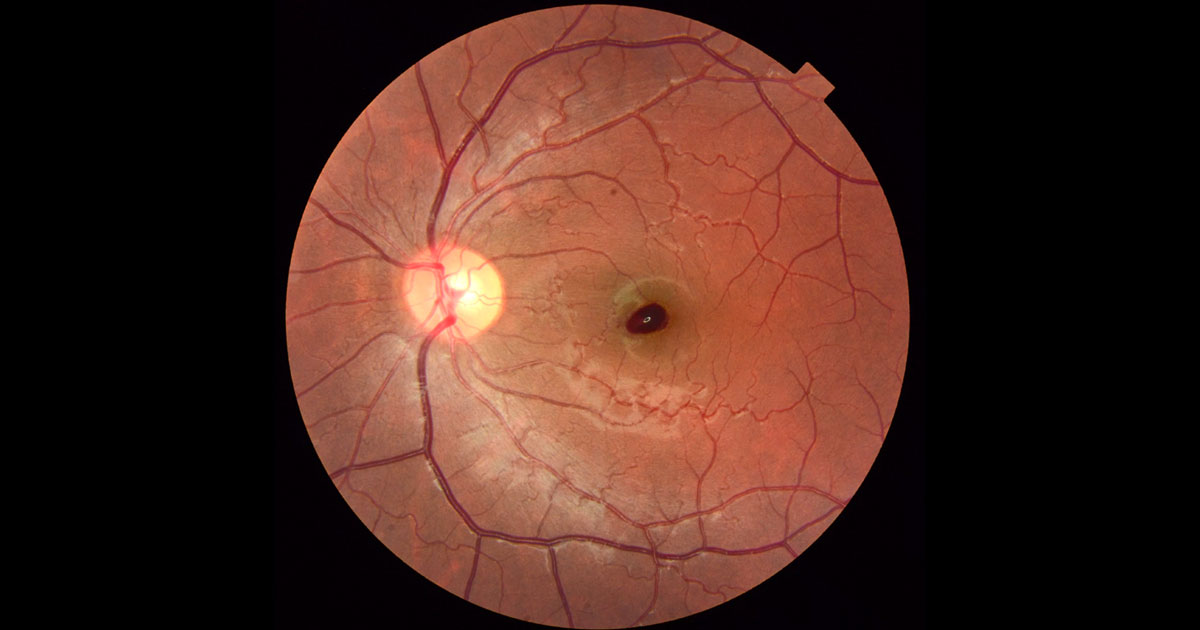
A 21-year-old female was referred with a dark patch at her left macula.
Tags: painless vision loss, scotoma, choroidal neovascular membrane, valsalva retinopathy
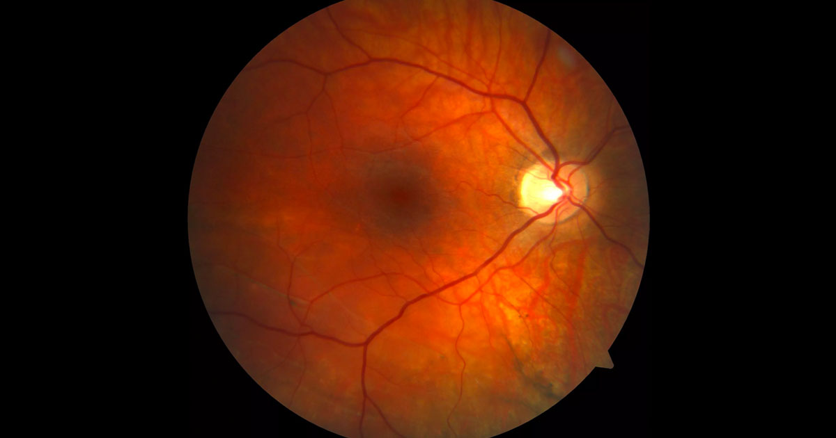
A 28-year-old male was referred with incidental retinal findings in his right eye.
Tags: myopia, retinal detachment
A 67-year-old female was referred with reduced vision.
Tags: reduced vision, bitemporal hemianopia, pituitary tumour, suprasellar meningioma
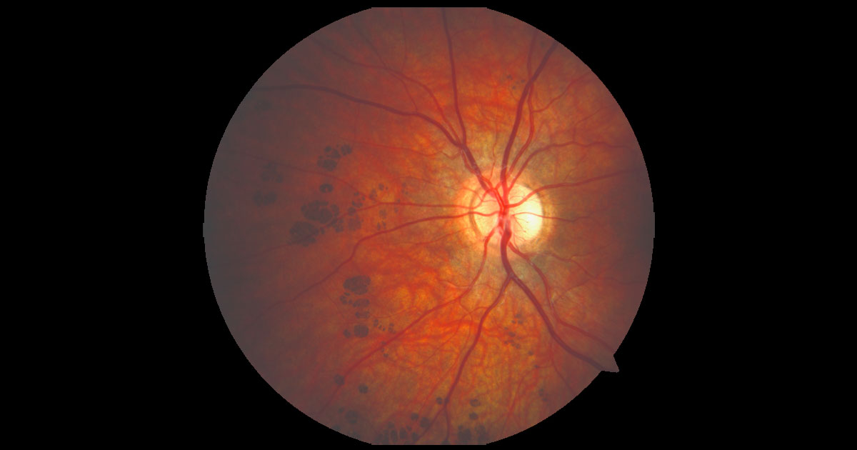
A 65-year-old Caucasian male was referred with unusual fundal lesions.
Tags: fundal lesions, subretinal nodules, sclerochoroidal calcification, granuloma
A 35-year-old lady was referred by her optometrist with diabetic retinopathy.
Tags: diabetic retinopathy, neovascularization, macula oedema, vision loss
A 14-year-old boy was referred by his optometrist with unusual areas of fundus pigmentation in his left eye.
Tags: fundus pigmentation, chrpe, familial adenomatous polyposis, vision loss
A 67-year-old male was referred with a 1 day history of painless inferior scotoma in his right vision.
Tags: scotoma, retinal pallor, branch retinal artery occlusion, cilioretinal artery occlusion
A 51-year-old high myope was referred with reduced vision in her left eye.
Tags: reduced vision, myopia, posterior staphyloma, myopic foveoschisis
A 31-year-old female was referred with floaters and reduced vision.
Tags: reduced vision, floaters, toxoplasma chorioretinitis, sarcoidosis
Have a question? Call one of our clinics today.
© 2019-2024 Eye Specialists Centre | Privacy Policy | Disclaimer | Website design: ![]()