Case 43
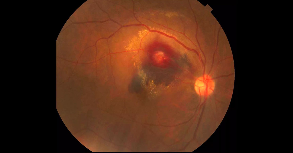
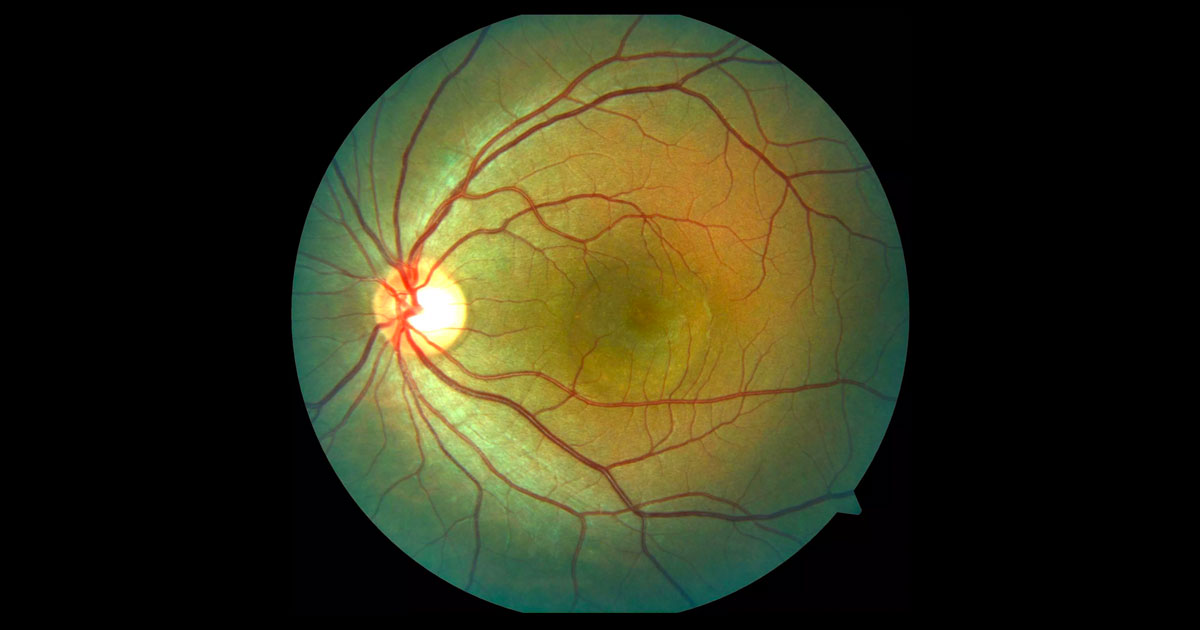
A 72-year-old woman was referred with acute painless vision loss in her right eye.
Here you’ll find interesting cases of eye conditions along with news and developments in the ophthalmology world.
Cases are presented as an initial image with history and examination. Health practitioners are encouraged to deduce the condition, before further investigations, diagnosis and management are presented.
We hope you find it as educational, informative and exciting as we do!
Click here to view our newsletter privacy notice.
The information provided during signup is used by Eye Specialists Centre to send newsletters using the cloud-based software, Mailchimp. We do not disclose or share your personal data with other third party without your consent, or unless it is required by law. If you have any concerns about your privacy, please do not hesitate to ask.
A 72-year-old woman was referred with acute painless vision loss in her right eye.
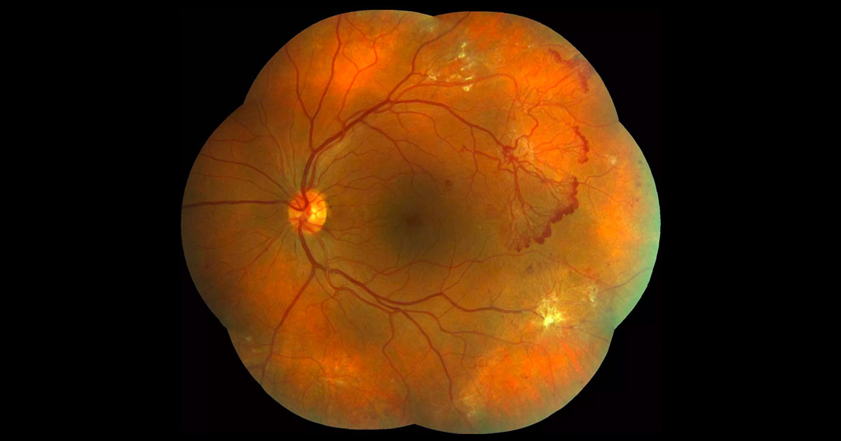
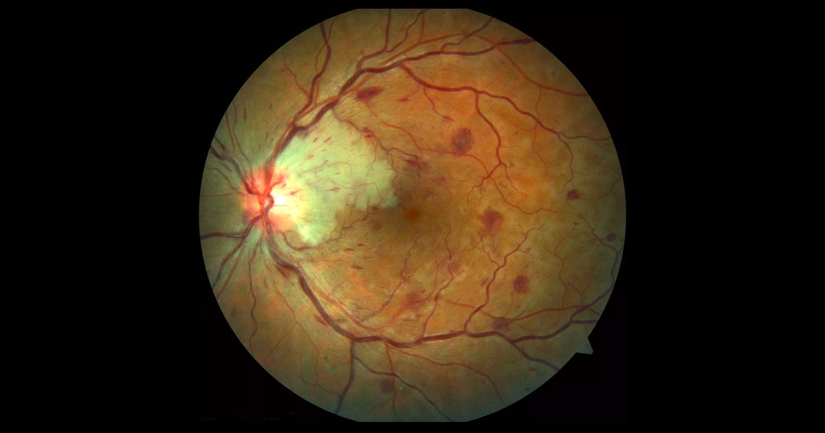
A 35-year-old lady was referred by her optometrist with diabetic retinopathy.
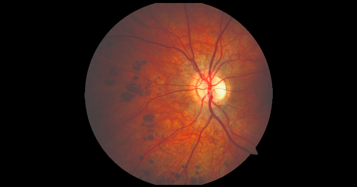
A 14-year-old boy was referred by his optometrist with unusual areas of fundus pigmentation in his left eye.
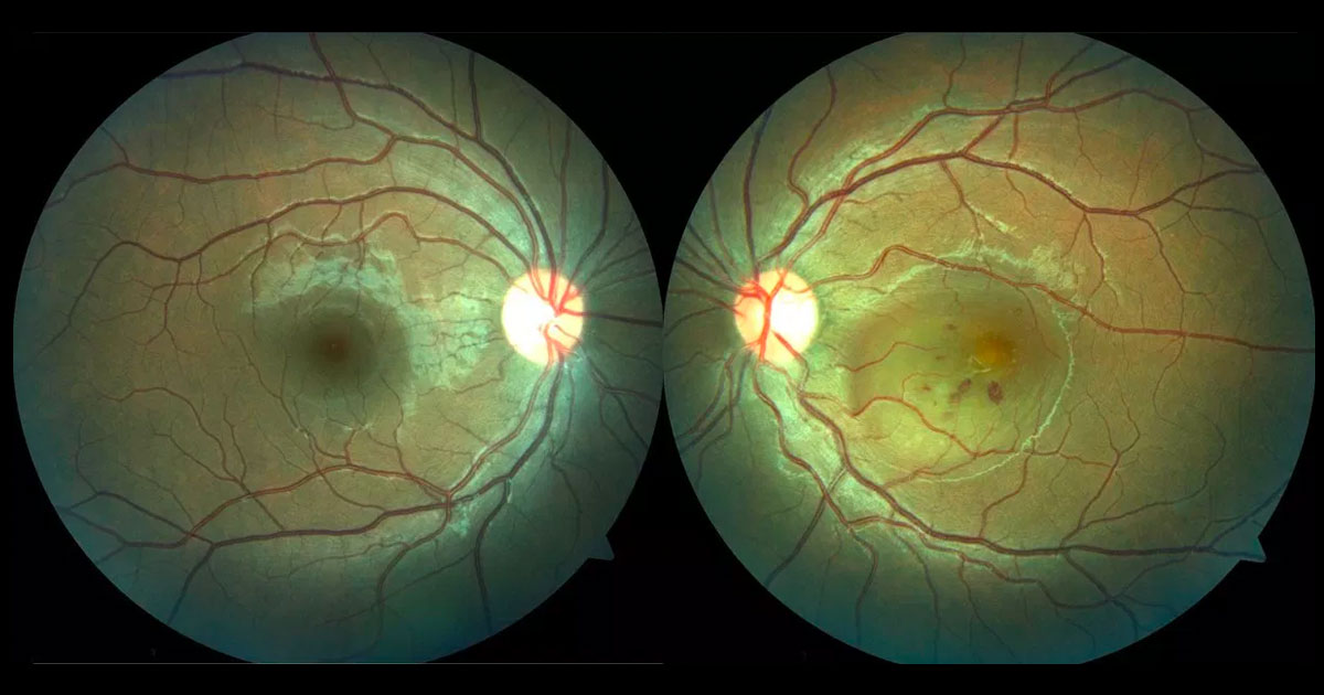
A 20-year-old man was referred with a central scotoma in his left eye.
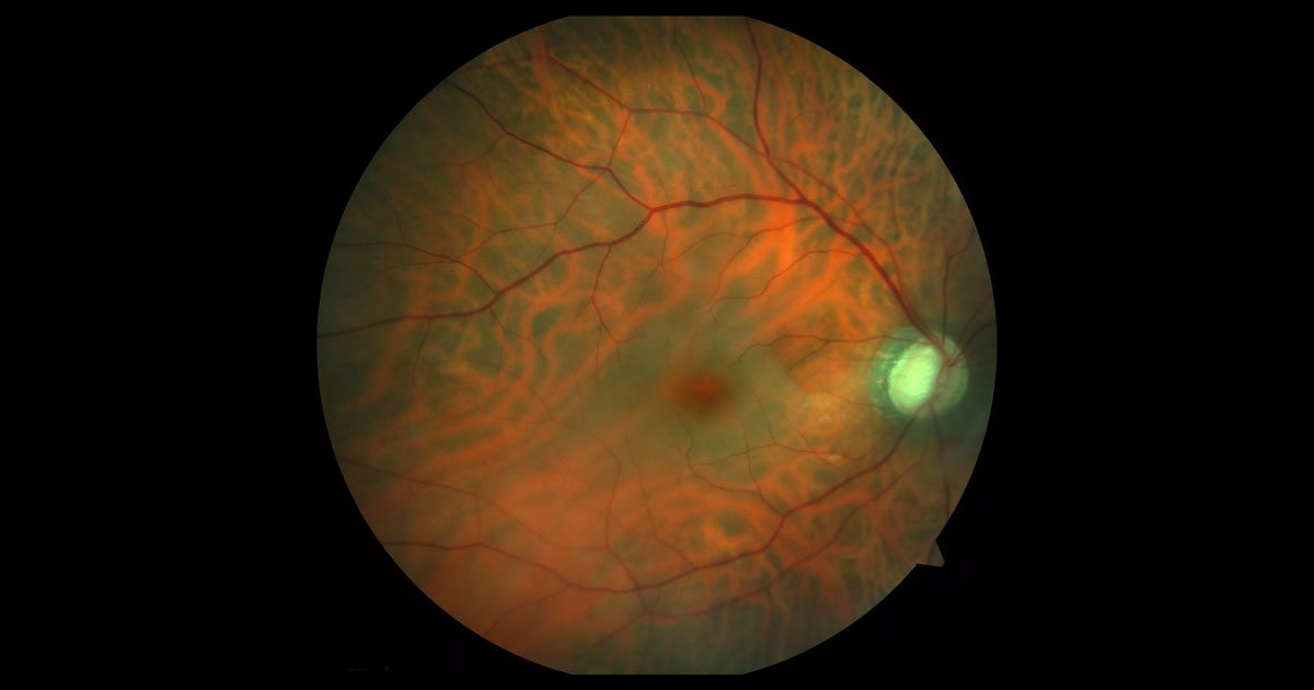
Figure 1. Colour photograph of the right fundus at presentation demonstrates retinal pallor with a cherry red spot at the macula. The cup disc ratio is enlarged.
A 68-year old man was referred with profound vision loss one day following cataract surgery.
A 22-year-old man presented with a “blob” in the centre of his right vision.
A 39-year-old man was referred with gradual loss of vision in both eyes noted over the last six months.
A 76-year-old man was referred with acute painless visual loss in his right eye.
A 32-year-old man was referred with blurring of his left central vision.
A 37-year-old lady was referred with acute painless left vision loss
A 32-year-old nurse was referred complaining of distorted central vision in her right eye.
Have a question? Call one of our clinics today.
© 2019- Eye Specialists Centre | Privacy Policy | Disclaimer | Website design: ![]()