Case 56
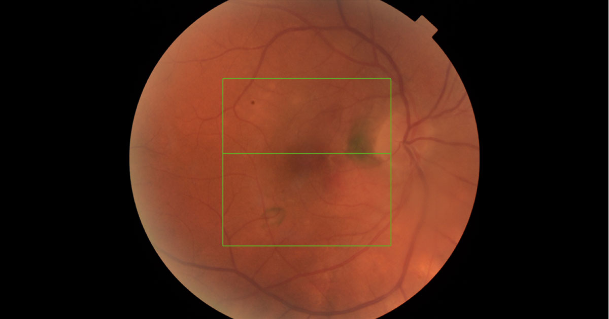
Figure 1.
Author: Amy Pai Editor: Adrian Fung
A 66-year-old Caucasian lady presented to her local ophthalmologist complaining of a 2 months history of blurry vision in her right eye.
Case history
A 66-year-old Caucasian lady presented to her local ophthalmologist complaining of a 2 months history of blurry vision in her right eye. She was diagnosed with right peri-papillary CNVM a year ago and treated with six Bevacizumab injections to date. They were started at one monthly intervals and now given on a PRN basis. She was referred to us for a second opinion and the treating ophthalmologist has noticed bilateral vitreous cells recently. She denied any other previous eye issues or operations and only takes an antihypertensive normally. She denied smoking and there is no family history of ocular problems. She normally wears glasses for reading.
Visual acuity was 6/18 (pinhole no improvement) in the right eye (OD) and 6/6 in the left eye (OS). Mild nuclear sclerotic cataracts were present in both eyes. There was no anterior chamber cells, flare, or posterior synechiae. Pupil reaction was within normal with no RAPD present. Pigmentary changes were noted in the right peri-papillary region and there was also an area of pigmentary change inferotemporal to the fovea. (Figure 1 shows the right fundus, the view was hazy due to vitritis). There were also irregularities in the retinal vessels, suggestive of vasculitis. The optic disc was full in appearance but with clear margins. Figure 2 shows the left fundus, there were also some RPE changes within the posterior pole, as well as similar vessel appearance suggestive of retinal vasculitis. The view is also hazy due to vitreous activity.
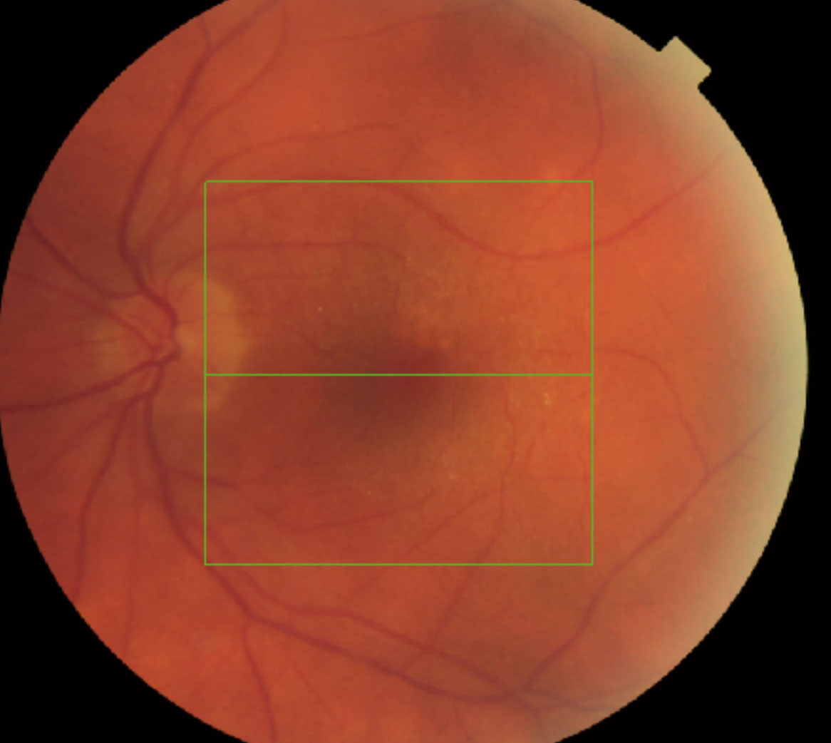
Figure 2. RPE changes in the macular region with irregular vasculature suggestive of vasculitis.
What is your diagnosis?
Click to reveal answer
Differential diagnosis
The differential diagnosis includes:
- Posterior Uveitis with right inflammatory CNVM
- ARMD with right CNVM
- Endophthalmitis post AntiVEGF injections
Additional history, examination and investigations
OCT scans, FFA and ICG were performed in our clinic and a clinical diagnosis of Birdshot Chorioretinopathy with right inflammatory CNVM was made. We sent off patient’s blood samples for HLA A29 to confirm the diagnosis, as well as other potential causes of posterior uveitis including TB Quantiferon, ACE, and Syphilis VDRL. A full blood count, liver and renal function tests were performed. A Chest X ray was also requested to exclude pulmonary lesions. HLA A29 returned positive, whereas all the other investigations were negative.
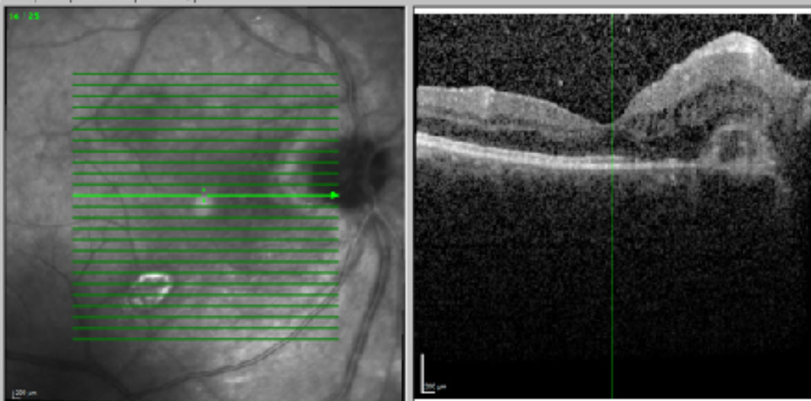
Figure 3. Right optical coherence tomography through the macula. There is a subretinal hyper reflective lesion close to the optic disc with some subretinal and intraretinal fluid. The fovea was just spared. There are vitreous cells present.
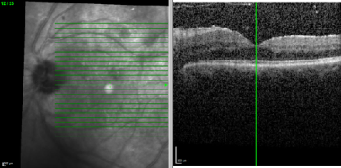
Figure 4. Left optical coherence tomography through the macula. There is retinal thickening with few intraretinal cystic changes close to the disc. There are also vitreous cells present.
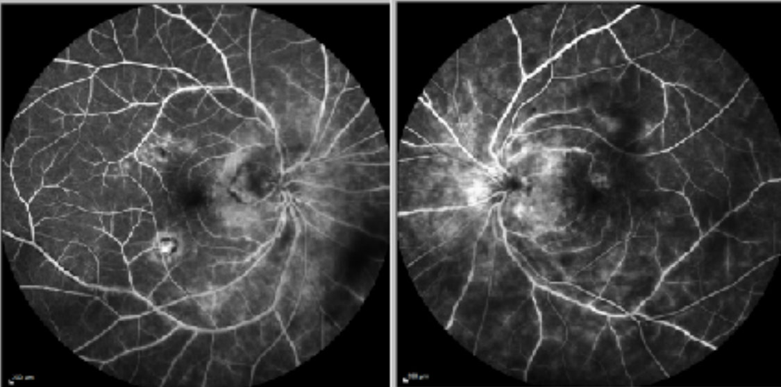
Figure 5. Fundus fluorescein angiography (FFA) confirming bilateral vasculitis, some disc leakage and right CNVM peripapillary and inferotemporal to the fovea.
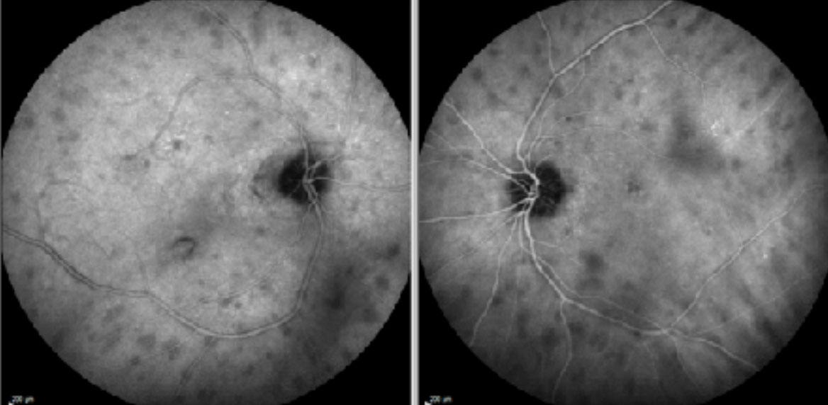
Figure 6. Late phases of Indocyanine Green Angiography (ICGA), showing multiple hypo-fluorescent choroidal lesions typically seen in Birdshot Chorioretinopathy.
DIAGNOSIS
Bilateral Birdshot Chorioretinopathy with right inflammatory CNVM.
Clinical course
Given the clinical findings, we commenced the patient on a course of oral Prednisolone with introduction a steroid sparing agent early to control the inflammation. She was initially started on oral Prednisolone 60mg daily po (body weight 67kg), and currently on a slow tapering course. Tacrolimus was introduced after trialling and failing Mycophenolate. The Tacrolimus level was monitored with serum trough levels and dose adjusted according to her clinical progression. The patient continued to receive right Bevacizumab injections on a PRN basis, and has not required many more injections since commencing systemic treatment for Birdshot. Vitreous activity has also decreased since commencing systemic treatment. Her right vision has improved to 6/9 at the last visit with no complaint of seeing floaters. She is under regular 3 monthly review at present.
Discussion
Birdshot chorioretinopathy, or commonly known as Birdshot, is a chronic bilateral posterior uveitis that involves inflammation of the choroid and retinal vessels. Patients commonly present complaining of reduced vision and floaters, and fundus examination may reveal multiple hypopigmented lesions, clustered and radiating out from the optic discs. They may present early with vitritis and vasculitis,(1) and later in the disease with cystoid macular oedema (CMO), and retinal/choroidal atrophy. They are almost never seen with anterior chamber inflammation, keratic precipitates, or posterior synechiae.(2)
Birdshot is primarily associated with Caucasian females, aged between 40-60 years of age. It has the strongest correlation with HLA A29, with 80-98% of patients being positive, versus 7% in the general population.(3)
As the disease progresses, Birdshot may present with CMO as the main cause of reduced vision (84% vs 30% in other forms of uveitis),(1) RPE changes or atrophy, optic disc atrophy and inflammatory CNVMs. Therefore, regular OCT scans, visual field tests, fundus photography, fundus autofluorescence, colour vision, FFAs and ICGs are important diagnostic and monitoring tests. Electrophysiology tests are also helpful in monitoring the disease progression.(4)
About 20% of Birdshot may achieve remission without treatment, but majority of patients will require long term immunomodulation due to recurrent attacks. Oral or local steroids may be used for acute exacerbations, and chronic disease will often require long term immunosuppressants such as Mycophenolate, Cyclosporine, or Tacrolimus. These medications will require regular blood tests to monitor for side effects. Although CNVMs are rare complications of birdshot (less than 6% in reported cases),(3) often combination treatment of AntiVEGF injections and systemic anti-inflammatories are required to prevent permanent visual loss.(5)
TAKE HOME POINTS
- Birdshot Chorioretinopathy is a chronic, usually bilateral cause of posterior uveitis
involving the choroid and retinal vessels. - Birdshot normally affects females, in their 40-60’s, of Caucasian ethnicity.
- It has a strong correlation with HLA A29.
- It rarely presents with anterior chamber inflammation.
- CMO, RPE changes/atrophy, optic disc atrophy and CNVMs are potential complications of Birdshot.
- Birdshot often requires long term systemic immunosuppression to prevent and treat disease progression and exacerbations. Although CNVMs are rare complications, but will require AntiVEGF injections in addition to systemic treatment.
REFERENCES