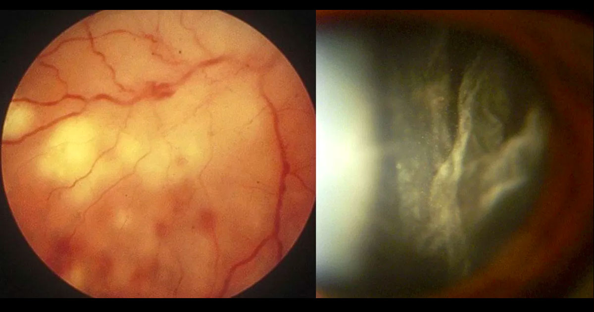
Figure 1. Colour fundus photograph of the right eye demonstrates white necrotic retinitis with intraretinal haemorrhages (left image). A moderate vitritis was present (right image).
A 54-year-old male was referred with reduced vision in his right eye.
A 54-year-old male was referred from his optometrist with a 2 day history of blurring of vision in his right eye associated with floaters.
Best corrected visual acuities were 6/18 in the right eye (OD) and 6/60 in the left eye (OS). Intraocular pressures were 15 in both eyes. Examination of the anterior segments revealed 2+ cells in the right eye and a quiet left eye. Examination of the posterior segments revealed vitritis associated with an area of retinitis with surrounding intraretinal haemorrhages in the right eye (Figure 1), and geographic atrophy in the left eye with no active inflammation.
The differential diagnosis of retinitis associated with panuveitis includes:
- Bacterial endophthalmitis
- Toxoplasma chorioretinitis
- Acute retinal necrosis (HSV, VZV)
- CMV retinitis (not usually associated with vitritis)
- Syphilis
- Sarcoidosis
- Tuberculosis
- Behcet’s disease
- Lymphoma
The patient was otherwise healthy with no significant past history. He was immediately referred to hospital for a vitreous biopsy. PCR testing was positive for Herpes Simplex Virus-1 (HSV-1). Other blood tests were normal.
DIAGNOSIS
Acute retinal necrosis secondary to HSV-1 infection.
The patient was admitted to hospital and received repeat intravitreal injections of the anti-viral Foscarnet over a one week period. He received intra-venous administration of the antiviral acyclovir. Oral steroids were commenced after a week. There was steady improvement in the retinitis and vitritis and the patient was discharged 10 days later on oral antiviral agents and a tapering course of oral steroid. He is currently being monitored closely for the possible development of retinal detachment.
Acute retinal necrosis (ARN) is a typically unilateral panuveitis characterised by one or more foci of retinal necrosis with discrete borders. There is usually significant associated anterior uveitis and vitritis, and often occlusive retinal arteritis. Without treatment the retinal lesions progress rapidly, and there is often bilateral involvement and significant visual morbidity.
While varicella zoster virus (VZV) or one of the herpes simplex viruses (HSV-1 or HSV-2) is most often the cause of ARN, cytomegalovirus (CMV) occasionally causes the condition.(1) In addition, rare cases of Epstein-Barr virus-associated retinitis and panuveitis have occurred.
Acute retinal necrosis syndrome is uncommon. Two recently published nationwide surveys from the United Kingdom estimated the incidence of ARN at one case per 2 million people per year.(2,3) It typically presents with eye redness, periorbital pain, photophobia, and vision loss. Anterior-segment findings include episcleritis, scleritis, keratitis, and/or anterior chamber inflammation, which may be either nongranulomatous or granulomatous.
Posterior-segment findings include vitreous inflammation, one or more areas of full-thickness necrotizing retinitis, and occlusive arteritis. Signs of optic neuropathy may be present when the optic disc is involved. In approximately one-third of patients, the fellow eye may become involved, typically within six weeks of initial presentation.(4)
The wider availability of polymerase chain reaction (PCR) testing of vitreous fluid has greatly assisted diagnosis.(5) Such testing is highly sensitive for the detection of viral DNA even in small volume vitreous samples, although sensitivity can fall dramatically if antiviral therapy has already been initiated.
Intravitreal and systemic antiviral therapy forms the mainstay of treatment. Foscarnet is the preferred intravitreal agent due to its broad spectrum of viral coverage and low incidence of viral resistance, whilst systemic treatment can be given in the form of intravenous acyclovir (requiring hospital admission), or oral valacyclovir.(6) There is typically a prompt response to treatment and systemic steroid may be added later to assist with resolution of the associated vitritis. Oral antiviral treatment is usually continued for at least 3 months.(7)
Some clinicians advocate for the use of prophylactic laser photocoagulation around the area of retinitis, in order to reduce the risk of retinal detachment. However this remains controversial and there is no conclusive evidence supporting its use(8).
Retinal detachment can occur in up to three quarters of patients and presents a significant management challenge and threat to vision.(1) There is often both a rhegmatogenous component, due to retinal necrosis, and a tractional component. Long acting gas or silicone oil tamponade may be required.
TAKE HOME POINTS
- Acute retinal necrosis (ARN) is a serious, rare panuveitis which most commonly occurs in otherwise healthy individuals.
- The classical lesion is a focal area of necrotising retinitis associated with vitritis.
VZV, HSV-1 and 2 are the most common causative viruses.
- PCR testing of vitreous fluid has significantly improved the accuracy of diagnosis.
- Intravitreal and systemic antiviral agents are the mainstay of therapy.
- Prompt recognition of this condition and referral can significantly improve outcomes.
REFERENCES
- Lau CH, Missotten T, Salzmann J, Lightman SL. Acute retinal necrosis features, management, and outcomes. Ophthalmology 2007;114:756-62.
- Cochrane TF, Silvestri G, McDowell C, Foot B, McAvoy CE. Acute retinal necrosis in the United Kingdom: results of a prospective surveillance study. Eye (Lond) 2012;26:370-7; quiz 8.
- Muthiah MN, Michaelides M, Child CS, Mitchell SM. Acute retinal necrosis: a national population-based study to assess the incidence, methods of diagnosis, treatment strategies and outcomes in the UK. Br J Ophthalmol 2007;91:1452-5.
- Fisher JP, Lewis ML, Blumenkranz M, et al. The acute retinal necrosis syndrome. Part 1: Clinical manifestations. Ophthalmology 1982;89:1309-16.
- Knox CM, Chandler D, Short GA, Margolis TP. Polymerase chain reaction-based assays of vitreous samples for the diagnosis of viral retinitis. Use in diagnostic dilemmas. Ophthalmology 1998;105:37-44; discussion -5.
- Wong R, Pavesio CE, Laidlaw DA, Williamson TH, Graham EM, Stanford MR. Acute retinal necrosis: the effects of intravitreal foscarnet and virus type on outcome. Ophthalmology 2010;117:556-60.
- Morse LS, Mizoguchi M. Diagnosis and management of viral retinitis in the acute retinal necrosis syndrome. Semin Ophthalmol 1995;10:28-41.
- Park JJ, Pavesio C. Prophylactic laser photocoagulation for acute retinal necrosis. Does it raise more questions than answers? Br J Ophthalmol 2008;92:1161-2.
Back
Tags: blurred vision, intraretinal haemorrhage, HSV, retinitis