Case 30
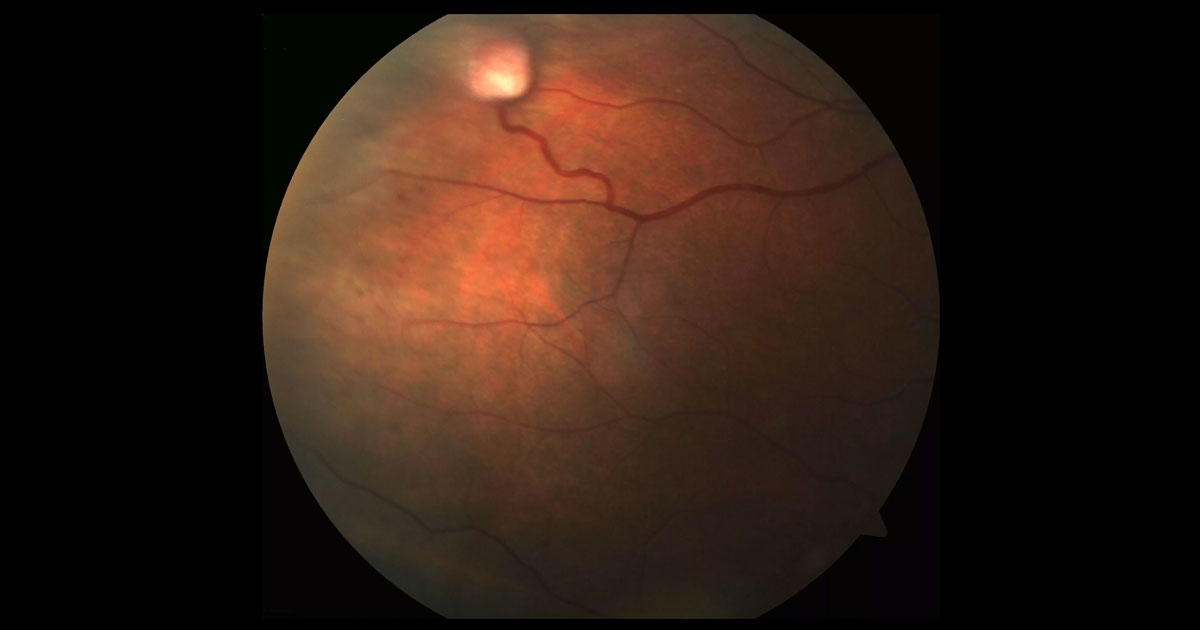
Figure 1. Colour fundus photograph of the right eye shows a raised white lesion in the supero-temporal periphery covered with fine vessels. There is a feeder arteriole and a dilated venous draining vessel.
Author: Dov Hersh Editor: Adrian Fung
A 33-year-old female was referred with a right supero-temporal retinal lesion.
Case history
A 33-year old-female was referred with a right supero-temporal retinal lesion. The left eye had undergone enucleation 10 years prior due to rubeotic glaucoma.
Visual acuity was 6/6 in the right eye and the intraocular pressure was 12mmHg. There was a well fitting prosthesis in the left socket. The lens and vitreous were clear. Right fundus examination showed a raised white lesion in the supero-temporal periphery measuring 2mm in width. This had a feeder arteriole and a dilated venous draining vessel (Figure 1).
What is your diagnosis?
Click to reveal answer
Differential diagnosis
The differential diagnosis for a raised peripheral vascular retinal lesion includes:
- Retinal capillary haemangioblastoma
- Coats disease
- Retinal arteriolar macroaneurysm
- Peripheral choroidal new vessel
- Vasoproliferative tumour
- Retinal cavernous haemangioma
- Granuloma with vasculitis
Additional history, examination and investigations
On further history the patient had a known diagnosis of von Hippel-Lindau disease. She undergone bilateral adrenalectomy in 2010 and was diagnosed with a cerebellar haemangioblastoma in 2013. The patient was being actively managed by physicians for her systemic conditions, but had not seen an ophthalmologist since 2007. The left eye had previously been enucleated due to debilitating pain secondary to total retinal detachment and neovascular glaucoma as a result of untreated retinal capillary haemangioblastomas.
Further family medial history was obtained. The patient’s father had been diagnosed with VHL, however no further medical history was known. The patient’s 12-year-old daughter tested positive for VHL, and had seen a paediatrician 2 years prior, but not an ophthalmologist.
DIAGNOSIS
Retinal capillary haemangioblastoma in association with von Hippel-Lindau disease (VHL).
Clinical course
Given the propensity for retinal capillary haemangioblastoma associated with VHL to progress, argon laser photocoagulation to the feeder arteriole, lesion and draining venule was applied (Figure 2). Review 2 weeks post-laser revealed regression of the retinal capillary haemangioblastoma and chorioretinal atrophy along the treated feeding arteriole and draining venule (figure 3).
The patient continues to be monitored on a regular basis for development of further haemangioblastomas. The patient’s 12-year-old daughter was invited to come for review. Visual acuity was 6/6 in both eyes, and intraocular pressures were 14mmHg (right eye) and 13 mmHg (left eye). Lens examination was normal and the vitreous was clear. Fundus examination of the left eye revealed a retinal capillary haemangioblastoma emanating from the optic nerve head (Figure 4). The remainder of the retinal examination in both eyes was normal, with no further retinal capillary haemangioblastomas detected.
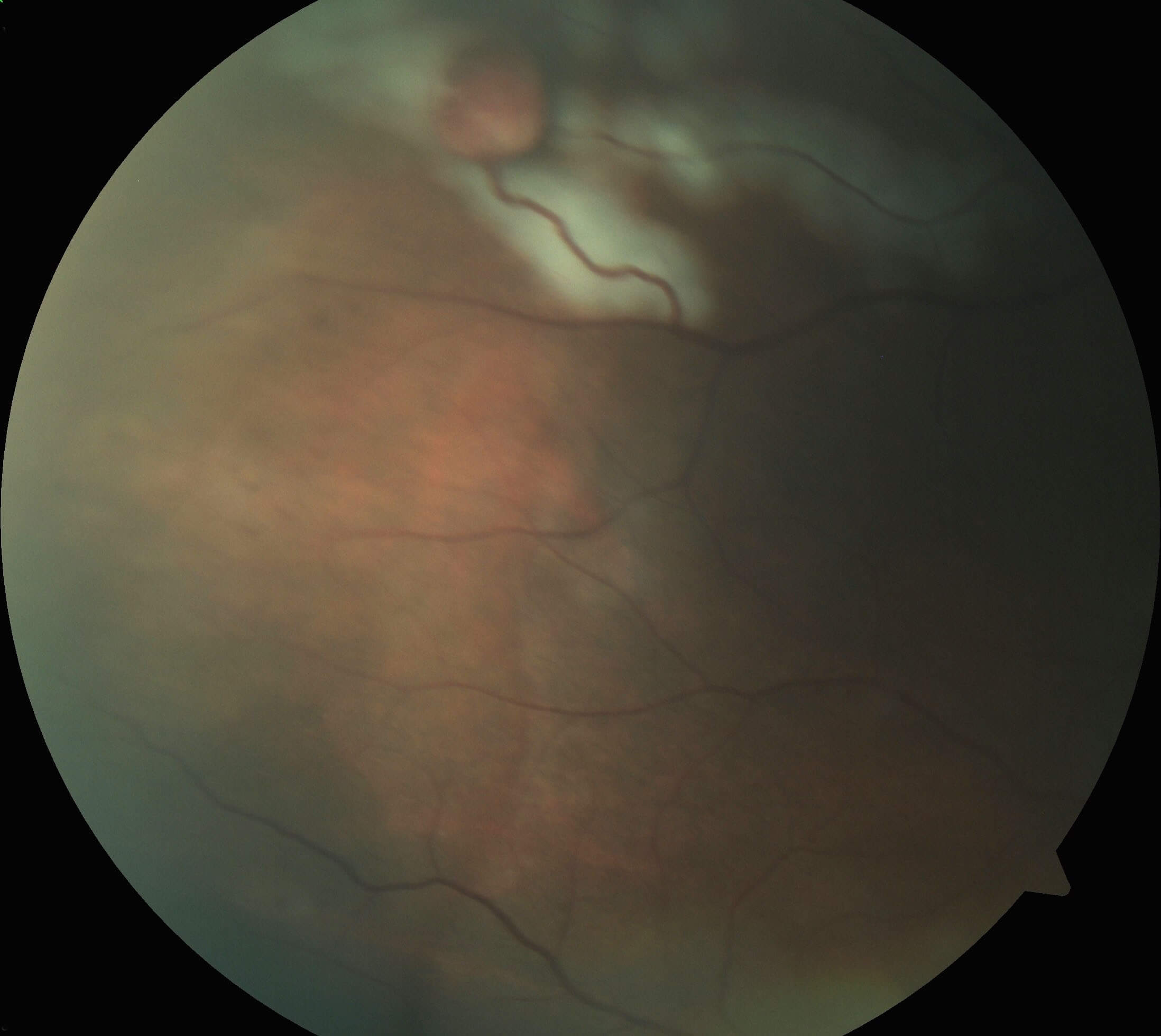
Figure 2. Colour fundus photograph immediately following argon laser photocoagulation. The laser marks are seen in white.
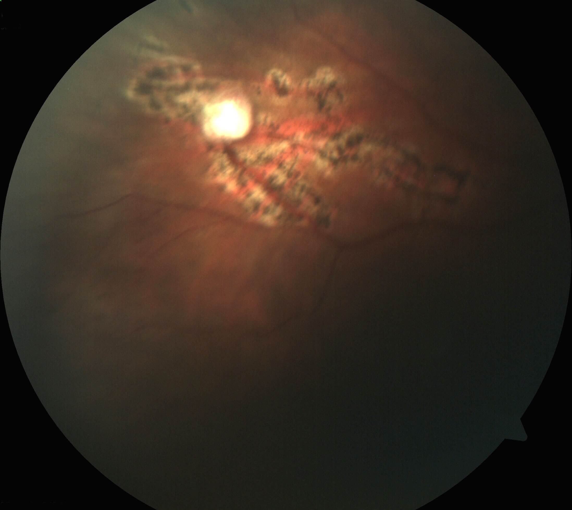
Figure 3. Colour fundus photograph two weeks post laser photocoagulation.
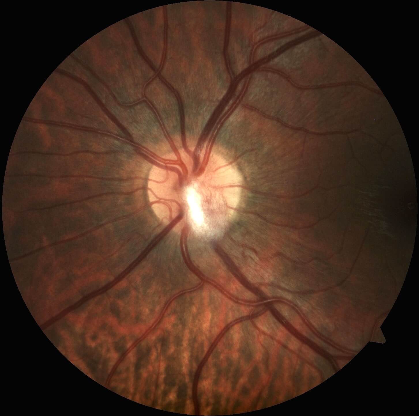
Figure 4. Colour fundus photography of the left eye in the patient’s daughter demonstrates a retinal capillary haemangioblastoma emanating from the optic nerve head.
Discussion
von Hippel-Lindau disease (VHL) is a highly penetrant, autosomal dominant, multisystem cancer syndrome caused by a germline mutation of the VHL tumour suppressor gene on the short arm of chromosome 3 (3p25–26). About 20% of cases are new mutations, with no family history of the disease. Patients with VHL are at increased risk of developing retinal and central nervous system hemangioblastomas, renal cell carcinomas, pheochromocytomas, neuroendocrine tumors, cysts of the pancreas and endolymphatic sac tumors.(1)
Patient care necessitates a multi-disciplinary approach to regular and lifelong surveillance for ocular and systemic tumours. Well-documented screening protocols are available.(2) It is generally accepted that patients with VHL should have yearly retinal exams from the age of 5 years to screen for RCH. Highly specific and sensitive genetic testing is available to detect patients with the VHL mutation. In addition, antenatal testing is possible.(3)
Retinal capillary haemangioblastomas (RCH) are present in up to 70% of individuals with VHL, and are the most frequent and often the earliest manifestation of the disease. By definition, patients can be diagnosed with VHL if they have:
- More than 1 RCH
- 1 RCH and an associated visceral lesion
- 1 RCH with a family history of VHL.(1)
The mean age at diagnosis of RCHs in VHL disease is approximately 25 years, although they can present in the early teens as occurred in our patient. In patients with VHL, RCHs often develop bilaterally, with new lesions developing over time. The lesions are red-colored, highly vascular with a characteristic feeder artery and draining venule. Most commonly the lesions are located in the mid-periphery, however 15% of RCHs occur within one-disc diameter of the optic nerve head.(4)
Although small lesions may remain stable for years, most RCHs associated with VHL tend to progress. Complications including massive exudation, retinal detachment, macula oedema, uveitis, glaucoma and phthisis may occur.(4) Treatment of RCHs is aimed at tumour ablation in order to prevent further growth and complications. Treatment options for peripheral lesions include laser photocoagulation, cryotherapy, radiation and photodynamic therapy. Laser photocoagulation is used to treat small RCHs in the posterior retina. Ideally the feeder artery is treated first followed by the tumour’s surface.(5) Cryotherapy is useful for anteriorly located lesions not amenable to laser photocoagulation, or lesions with subretinal fluid.
Lesions of the optic nerve head (juxtapapillary) present a particular treatment challenge. If left untreated, these lesions tend to increase slowly in size over time and ultimately cause exudation and visual loss. Unfortunately, active ablative treatment may cause direct damage to the optic nerve resulting in visual loss. Modalities including photodynamic therapy, proton beam radiotherapy and anti-VEGF agents have been trialed with limited success.(6)
TAKE HOME POINTS
- Retinal capillary haemangioblastomas are vascular tumours that usually occur in the retinal periphery with a feeding arteriole and draining venule.
- Retinal capillary haemangioblastomas may be isolated lesions, or associated with von Hippel-Lindau disease.
- von Hippel-Lindau disease is an autosomal dominant condition associated with haemangioblastomas of the retina and brain, as well as visceral tumours.
- Retinal capillary haemangioblastomas associated with von Hippel-Lindau tend to progress and early ablative treatment with laser photocoagulation or cryotherapy should be considered to avoid complications.
- Retinal capillary haemangioblastomas of the optic nerve head (juxtapapillary) present a particular management challenge.
REFERENCES